
Bone Tumour Treatment in Gurgaon
Bone tumours can range from harmless benign growths to aggressive cancers such as osteosarcoma, Ewing’s sarcoma, and chondrosarcoma, making early recognition and expert care essential. Persistent bone pain, unexplained swelling, or a lump that doesn’t go away are warning signs that should never be ignored. Seeking timely Bone Tumour Treatment in Gurgaon ensures accurate diagnosis and access to advanced care options.
Dr. Ramkinkar Jha, Director, Department of Orthopaedics at CK Birla Hospital, Gurugram, is one of the most experienced orthopaedic oncologists in Delhi NCR, having managed complex bone and soft-tissue tumour cases across all age groups, from adolescents with primary bone cancers to adults with metastatic bone disease. With 20+ years of surgical experience and fellowship training from the UK, Singapore, and Hong Kong, Dr. Jha combines advanced imaging-guided diagnosis with limb-salvage techniques that prioritise both cancer clearance and long-term function, avoiding amputation wherever oncologically safe.
Book a Consultation with Dr. Ramkinkar Jha, Leading Orthopaedic Surgeon in Gurgaon
What Is a Bone Tumour?
A bone tumour is an abnormal mass of tissue that grows inside or around a bone. It can be benign (non-cancerous) or malignant (cancerous). Benign tumours don’t spread to other parts of the body, but they can still cause pain, weaken the bone, and need treatment. Malignant bone tumours are far more serious and require prompt, specialist-led care.
The good news is that most bone tumours are benign. Primary bone cancer accounts for roughly 0.2% of all malignancies worldwide, making it relatively rare. But rare doesn’t mean it can be ignored. When a malignant tumour is present, early diagnosis makes a real difference to outcomes.
There are two broad categories:
- Primary bone tumours, starting in the bone itself.
- Secondary bone tumours, also called metastatic bone disease, occur when cancer from another organ (such as the breast, prostate, or lung) spreads to the bones.
Each requires a different treatment approach, which is why an accurate diagnosis comes first.
Types of Bone Tumours
There are several bone tumour types, each with different behaviour, age group patterns, and treatment needs. The most common primary malignant types are osteosarcoma, Ewing’s sarcoma, and chondrosarcoma, while benign tumours include giant cell tumours, enchondromas, and osteoid osteomas.
Here’s a quick overview of the main types:
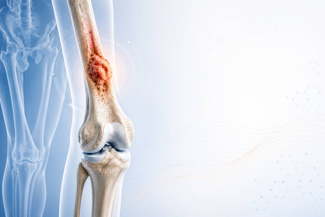
- Osteosarcoma: The most common primary malignant bone tumour, accounting for roughly 20% of all primary bone cancers. It most often affects teenagers and young adults, typically developing near the knee or shoulder. It grows fast and needs aggressive treatment.
- Ewing’s Sarcoma: The second most common primary bone cancer in children and adolescents, usually affecting those aged 4 to 15 years. Unlike osteosarcoma, it can start inside the bone or in the surrounding soft tissue. It spreads quickly and often presents with fever, fatigue, and pain together, which can lead to confusion with infections.
- Chondrosarcoma: A cancer of cartilage cells. It accounts for more than 40% of adult bone cancers and is most common in people over 40. It tends to grow more slowly than osteosarcoma and is less responsive to chemotherapy, making surgery the main treatment.
- Giant Cell Tumour of Bone: Usually benign but locally aggressive. It can destroy bone and sometimes recurs after treatment. Rarely, it can become malignant.
- Benign Tumours (Enchondroma, Osteoid Osteoma, Aneurysmal Bone Cyst): These are non-cancerous growths that may cause pain, deformity, or pathological fractures if untreated, but don’t spread to other organs.
- Metastatic Bone Disease: When cancers of the breast, prostate, lung, kidney, or thyroid spread to bones. This is the most common bone tumour seen in adults over 40 and requires a completely different treatment strategy from primary bone tumours.
Bone Tumour Risk Factors and Warning Signs
Bone pain that worsens at night, a visible or palpable bone lump, swelling around a joint, or a fracture that occurs with minimal force are the four symptoms that should prompt immediate orthopaedic evaluation. Don’t wait to see if they resolve on their own.
A systematic review of 1,452 paediatric bone tumour patients found that pain was the most prevalent symptom, present in 64% of cases, followed by swelling in 22%. The problem is that these symptoms are easy to attribute to other causes, such as sports injuries, arthritis, or growing pains. That mismatch leads to delays in diagnosis that can affect outcomes.
Key warning signs include:
- Persistent bone pain, especially pain that gets worse at night or doesn’t respond to standard pain relief
- A lump or swelling over a bone or joint, whether painful or painless
- A pathological fracture: a break that happens from a minor fall or no trauma at all
- Reduced movement in a nearby joint
- Unexplained fatigue, weight loss, or fever alongside bone pain (more common in Ewing’s sarcoma and metastatic disease)
Risk factors that raise the likelihood of a bone tumour:
- Family history of bone sarcoma or genetic syndromes like Li-Fraumeni syndrome
- Previous radiation therapy to a bone region
- Paget’s disease of bone, which can predispose to secondary bone sarcomas
- Hereditary retinoblastoma (associated with a significantly higher osteosarcoma risk)
- A history of prior chemotherapy (certain agents are linked to secondary osteosarcoma)
Most bone sarcomas, however, occur in people with no known risk factors. So if you have symptoms, don’t rule it out just because you have no family history.
How Is a Bone Tumour Diagnosed?
A bone tumour is diagnosed through a combination of imaging and tissue biopsy. Imaging tells you where the tumour is and how it looks. A biopsy tells you exactly what it is. Both are needed before any treatment plan can be made.
The diagnostic process typically follows this order:
- X-ray: The first step. An X-ray can reveal abnormal bone density, irregular margins, or a visible mass. It’s fast and accessible, but it doesn’t give the full picture.
- MRI (Magnetic Resonance Imaging): The most important imaging tool for local tumour staging. It shows the tumour’s exact size, how far it extends into soft tissue, and its relationship to nearby structures like nerves and blood vessels. MRI is essential for planning surgery.
- CT Scan: Used to assess how far the tumour has spread. A CT scan of the chest is routinely done to check for lung metastases, which is the most common site of spread for osteosarcoma.
- PET-CT Scan: Shows metabolic activity across the whole body. PET-CT is valuable for detecting distant metastases, particularly in staging Ewing’s sarcoma.
- Biopsy: The only definitive way to confirm what type of tumour is present. It’s critically important that the biopsy is performed by an experienced orthopaedic oncologist. An incorrectly placed biopsy can compromise limb salvage options later, making the site, technique, and timing of the biopsy a specialist decision.
Blood tests, including alkaline phosphatase and LDH levels, can provide supporting information, but they can’t diagnose a bone tumour on their own.
Bone Tumour Treatment Options in Gurgaon
Bone Tumour treatment depends on the type of tumour, its grade, stage, size, and location. Today’s approach to bone tumour treatment combines surgery, chemotherapy, radiotherapy, and rehabilitation in a coordinated plan. The era of routine amputation is over. Modern orthopaedic oncology treatment prioritises preserving the limb wherever oncologically safe to do so.
- Surgical Resection: The foundation of treatment for most bone tumours. The goal is to remove the tumour with a clear margin of healthy tissue around it (a “wide margin”), leaving no cancer cells behind.
- Limb Salvage Surgery: The gold standard for most primary malignant bone tumours of the arms and legs. Instead of removing the limb, the surgeon removes the tumour and reconstructs the bone using metal implants (endoprostheses), bone grafts, or a combination of both. This is discussed in more detail in the next section.
- Neoadjuvant Chemotherapy: For high-grade tumours like osteosarcoma and Ewing’s sarcoma, chemotherapy is given before surgery to shrink the tumour and treat any microscopic spread. The tumour’s response to this pre-operative chemotherapy is one of the strongest predictors of long-term survival.
- Adjuvant Chemotherapy and Radiotherapy: Given after surgery to reduce the risk of recurrence. Ewing’s sarcoma is particularly responsive to radiation therapy.
- Minimally Invasive Techniques: For selected benign tumours, curettage (scraping out the tumour) combined with bone grafting can be done through smaller incisions with faster recovery.
- Metastatic Bone Disease Management: When cancer has spread to the bones from another primary site, treatment is guided by the original cancer type and focuses on pain relief, fracture prevention, and quality-of-life improvement. Surgical stabilisation may be needed for bones at risk of fracture.
A 2024 study comparing surgical approaches in patients with osteosarcoma found that those who underwent limb salvage surgery had a five-year survival rate of 83.8%, compared to 62.5% in the amputation group. Functional scores and quality of life were also significantly better in the limb-salvage group.
For tumours in the pelvis or around the hip socket, reconstruction becomes especially complex. This is where pelvic tumour reconstruction expertise matters, because the anatomy is demanding and the margin for error is narrow.
What Is the Prognosis for Bone Tumours?
Bone tumours prognosis depends primarily on tumour type, stage at diagnosis, and whether the cancer has spread beyond its original site. When caught early and treated at a specialist centre, many patients achieve long-term remission and good functional recovery.
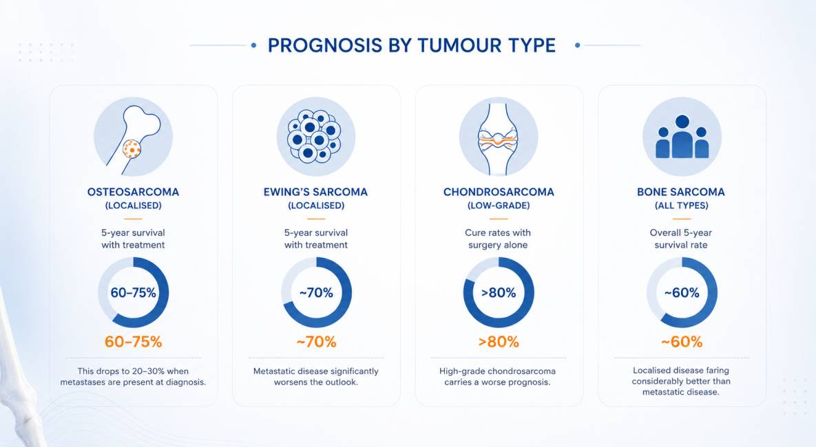
The most important factor within a patient’s control is timing. Bones that are evaluated early, before the tumour has grown large or spread, offer far more options. A structured diagnostic pathway and early specialist referral remain the most powerful tools available.
Struggling with Joint Pain, Sports Injury, or Arthritis?
Bone Tumour Treatment Cost in Gurgaon
The cost of bone tumour treatment in Gurgaon varies from one patient to another. It depends on several clinical and treatment-related factors, including:
- Type of bone tumour (benign or malignant)
- Tumour size, location, and stage
- Diagnostic tests required, such as MRI, CT scan, PET-CT, and biopsy
- Type and complexity of surgery, including limb salvage or reconstruction
- Need for chemotherapy or radiotherapy
- Implants or prostheses, if reconstruction is required
- Length of hospital stay and post-operative rehabilitation
After evaluating your reports and diagnosis, Dr. Ramkinkar Jha provides a personalised treatment plan with a transparent cost estimate. Patients travelling from other cities or overseas can also share their reports for an initial assessment before planning their visit.
Why Choose Dr Ramkinkar Jha for Bone Tumour Treatment in Gurgaon?
When the diagnosis involves a bone tumour, the surgeon’s experience with orthopaedic oncology is everything. This is not a condition where general orthopaedic care is sufficient. The biopsy technique, surgical margins, reconstruction approach, and coordination with medical oncology and radiation oncology all require specialist-level expertise.
What sets his approach apart:
- Specialist biopsy planning: The biopsy is done with the final surgery in mind, protecting limb-salvage options from the start.
- Tumour board discussions: Every malignant case is reviewed with a multidisciplinary team including medical oncologists, radiation oncologists, radiologists, and pathologists.
- Limb preservation focus: The default position is always to preserve the limb wherever it’s oncologically safe to do so.
- Global training, local availability: International fellowship experience applied in a patient-centric environment at one of Gurgaon’s leading hospitals.
- Transparent, empathetic communication: Complex diagnoses need clear explanations. Dr Ramkinkar Jha is known for taking the time to ensure patients and families understand every step.
You can learn more about Dr Ramkinkar Jha’s training and expertise on his profile page.
International Patients: Bone Tumour Treatment in Gurgaon
Gurugram is a recognised destination for international patients seeking bone tumour treatment in India. Gurgaon’s location in the Delhi NCR region, with direct access to Indira Gandhi International Airport, makes it straightforward to reach from South Asia, the Middle East, Africa, and beyond.
Dr Jha’s team routinely supports patients travelling from Bangladesh, Nepal, Afghanistan, Kenya, Nigeria, Uzbekistan, and the Gulf region for specialist orthopaedic oncology care.
What international patients can expect:
- Pre-arrival consultation via WhatsApp or video call to review existing reports
- Coordinated diagnostic and treatment scheduling to minimise time in-country
- Assistance with treatment summaries and discharge documentation
- Post-treatment follow-up by telemedicine
If you or someone in your family has been dealing with unexplained bone pain, a growing lump, or a recent scan that flagged something unusual, don’t put it off any longer.
Book a consultation with Dr Ramkinkar Jha in Gurugram.
Frequently Asked Questions
What complications can occur if a bone tumour is left untreated? expand_more
Untreated bone tumours, whether benign or malignant, carry significant risks. Early evaluation and treatment avoid these complications and preserve more options for the patient.
What tests are involved in the diagnosis of bone tumours? expand_more
Blood tests such as alkaline phosphatase and LDH levels may support the diagnosis but can't confirm it on their own.
What are the risk factors for developing a bone tumour? expand_more
Most bone tumours develop without a clear cause. Known risk factors include a family history of bone sarcoma, prior radiation therapy to a bone region, Paget's disease of bone, certain inherited conditions, and previous chemotherapy with certain drug types.
Can bone tumours be prevented? expand_more
Primary bone tumours, including osteosarcoma and Ewing's sarcoma, cannot currently be prevented because most arise without identifiable causes.
How do I book an appointment with Dr. Ramkinkar Jha? expand_more
You can book a consultation through the appointment section on this page. Early evaluation is recommended for better outcomes.

Dr Ramkinkar Jha's Medical Content Team
Dr. Ramkinkar Jha’s medical content team specialises in producing accurate, clear, and patient-focused orthopaedic content. With a strong foundation in clinical knowledge and expertise in technical writing and SEO, the team translates complex orthopaedic and musculoskeletal information into reliable, easy-to-understand resources. Their work helps patients make informed healthcare decisions while reflecting Dr. Jha’s commitment to high-quality, expert care in joint replacement, trauma, sports injuries, and advanced orthopaedic treatments.
This content is reviewed by Dr. Ramkinkar Jha
Related Blogs
Sarcoma Awareness Month: The Cancer Too Many People Have Never Heard Of
It usually starts small. A lump on the thigh that doesn’t hurt. A dull ache…
Read Article → WhatsApp