
MCL & LCL Injury Treatment in Gurgaon
MCL and LCL injuries range from mild sprains to complete tears causing knee instability. Treatment depends on grade and ligament involved – conservative care for most MCL injuries, surgical reconstruction for Grade III LCL tears, multi-ligament damage, or failed non-surgical management.
Book a Consultation with Dr. Ramkinkar Jha, Leading Orthopaedic Surgeon in Gurgaon
What Are the MCL and LCL?
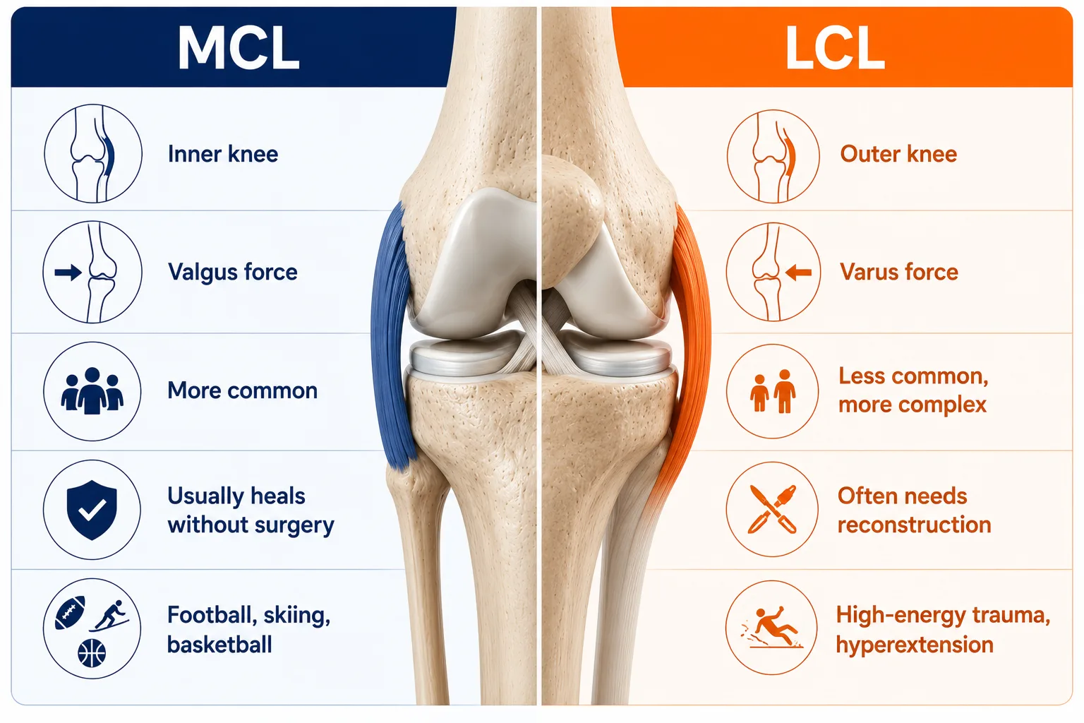
The knee is stabilised by four primary ligaments. Two of them – the Medial Collateral Ligament (MCL) and the Lateral Collateral Ligament (LCL) – run along the inner and outer sides of the knee respectively. Together, they form the collateral ligament complex, the joint’s primary defence against sideways (varus and valgus) forces.
- MCL: Runs along the inner (medial) side of the knee, connecting the femur (thighbone) to the tibia (shinbone). It resists inward buckling of the knee – the valgus force.
- LCL: Runs along the outer (lateral) side of the knee, connecting the femur to the fibula. It resists outward buckling – the varus force. The LCL is also a key component of the posterolateral corner (PLC), a complex of structures that controls rotational stability.
Both ligaments are graded by severity of injury:
- Grade I (Mild Sprain): The ligament is stretched but intact. The knee remains stable. Tenderness is localised and swelling is minimal.
- Grade II (Partial Tear): The ligament is partially torn. There is noticeable laxity on stress testing, though some fibres remain intact. Pain and swelling are more pronounced.
- Grade III (Complete Tear): The ligament is fully ruptured. The knee is frankly unstable. This grade frequently occurs alongside injuries to other structures – the ACL, PCL, or posterolateral corner.
Accurate grading is not academic. It directly determines whether conservative management will suffice or whether surgery is necessary.
Causes: How Do MCL and LCL Injuries Happen?
Collateral ligament injuries are overwhelmingly caused by contact and high-impact sport, though the mechanism differs between the two ligaments.
MCL injuries are the more common of the two. They typically result from:
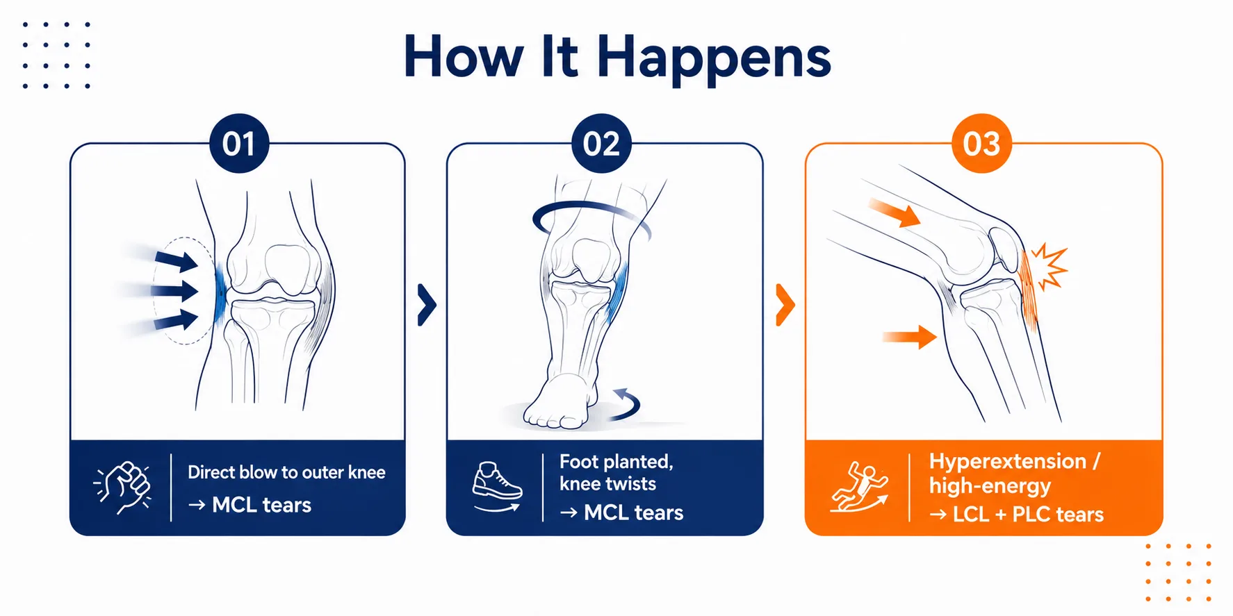
- A direct blow to the outer knee – a tackle in football or a collision in rugby that drives the knee inward (valgus force)
- Sudden twisting – pivoting with the foot planted, common in football, basketball, and martial arts
- Skiing falls – particularly when the ski catches and the knee twists inward
LCL injuries are less frequent but often more complex because the LCL is rarely injured in isolation. Common causes include:
- A direct blow to the inner knee – forcing the knee outward (varus force)
- Hyperextension injuries – the knee bending backward beyond its normal range, which can simultaneously damage the LCL and the posterolateral corner
- High-energy trauma – road traffic accidents and contact sport collisions that involve multi-directional force
In both cases, the force exceeds what the ligament can absorb elastically. The result is stretching, partial tearing, or complete rupture.
Recognising the Symptoms: MCL vs LCL Injury
The location of pain is the first and most reliable clue. Patients almost always point directly to the inner or outer knee – and they are usually right.
MCL Injury Symptoms
- Pain along the inner (medial) side of the knee, often sharp at the moment of injury and aching thereafter
- Swelling over the medial joint line, sometimes extending into the surrounding tissue
- Valgus instability – a sensation that the knee is giving way inward, particularly when changing direction or descending stairs
- Stiffness and difficulty fully bending or straightening the knee
- Bruising along the inner knee, appearing within 24–48 hours
Simple home test – Valgus Stress Test (MCL): Lie flat on your back with the knee slightly bent (about 20–30 degrees). Ask someone to gently apply an inward force at the ankle while stabilising the outer thigh. If this reproduces medial knee pain or reveals excessive inward movement compared to the other knee, MCL damage is likely. This is not a substitute for clinical assessment, but it is a useful early indicator.
LCL Injury Symptoms
- Pain along the outer (lateral) side of the knee, which can be sharp or deep depending on the grade
- Lateral instability – a feeling that the knee is buckling outward, especially on uneven ground
- Swelling over the lateral joint line, often less dramatic than MCL injuries because the LCL has a poorer blood supply
- Peroneal nerve symptoms – numbness, tingling, or weakness in the foot and outer lower leg. The common peroneal nerve runs close to the LCL and fibular head; it can be stretched or compressed in high-grade LCL injuries
- Difficulty walking in a straight line, with a tendency to compensate by rotating the hip
Simple home test – Varus Stress Test (LCL): In the same position as above, apply a gentle outward force at the ankle while stabilising the inner thigh. Pain at the outer knee or excessive outward movement compared to the uninjured side suggests LCL involvement. Again, clinical confirmation is essential.
Diagnosis of MCL and LCL Injuries
Because collateral ligament injuries range from minor sprains to complex multi-ligament tears, accurate diagnosis is non-negotiable. Dr. Ramkinkar Jha uses a structured, multi-step evaluation to grade the injury and identify any associated damage.
Physical Examination
- Valgus Stress Test (MCL): Performed at 0° and 30° of knee flexion. Laxity at 30° alone suggests an isolated MCL tear; laxity at both 0° and 30° raises concern for additional cruciate ligament involvement.
- Varus Stress Test (LCL): Applied at 0° and 30° to assess lateral laxity and posterolateral corner integrity.
- Dial Test: The patient’s feet are externally rotated at 30° and 90° of knee flexion. Increased external rotation at 30° (but not 90°) points to isolated posterolateral corner injury; increased rotation at both angles suggests combined PCL and PLC damage – a finding that significantly changes the surgical plan.
- Peroneal nerve assessment: Sensation and motor function in the foot are tested whenever LCL injury is suspected.
Imaging
- X-ray: The first step – to rule out fractures, particularly a Segond fracture (lateral capsule avulsion) or a fibular head avulsion, which are associated with LCL and PLC injuries.
- MRI (Magnetic Resonance Imaging): The gold standard for knee ligament injury treatment planning. MRI visualises the MCL and LCL fibres in detail, confirms the grade of the tear, and identifies concurrent meniscal, cruciate, or cartilage damage.
- CT scan: Ordered when a bony avulsion is suspected – for example, when the LCL pulls a fragment off the fibular head. CT defines the fragment size and displacement, which determines whether screw fixation is feasible.
Non-Surgical Treatment for MCL and LCL Injuries
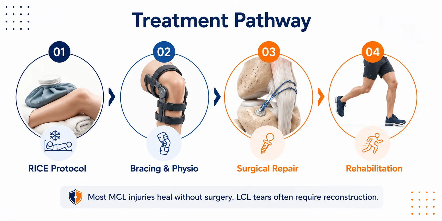
The good news: most MCL injuries and a significant proportion of Grade I–II LCL injuries heal without surgery. The key is protecting the ligament while it heals and rebuilding the dynamic stabilisers around the knee.
R.I.C.E Protocol for treatment for MCL and LCL Injuries (acute phase – first 48–72 hours):
- Rest: Avoid weight-bearing or use crutches to offload the knee
- Ice: Apply for 15–20 minutes every 2–3 hours to reduce swelling
- Compression: A firm bandage or compression sleeve limits effusion
- Elevation: Keep the leg raised above heart level to reduce oedema
Functional Bracing:
A hinged knee brace is applied to protect the healing ligament from further valgus or varus stress while allowing controlled movement. For MCL injuries, the brace is set to restrict the last few degrees of extension initially, then progressively opened as healing progresses.
Physiotherapy – the cornerstone of recovery:
- Quadriceps and hamstring strengthening to restore dynamic knee stability
- Hip abductor and external rotator strengthening to reduce valgus load on the MCL
- Proprioception and balance training to retrain neuromuscular control
- Gradual return to sport-specific movement patterns
Recovery timeline:
- Grade I: 4–6 weeks to full return to sport
- Grade II: 6–12 weeks, depending on compliance with bracing and physiotherapy
- Grade III (non-surgical candidates): 12 weeks or more; some isolated Grade III MCL tears in non-athletes can be managed conservatively, but this decision requires careful clinical judgement
MCL injury treatment in Gurgaon at this level – structured, progressive, outcome-focused – produces excellent results for the majority of patients when started promptly.
Surgical Treatment for MCL and LCL Injuries
Surgery is not the default for collateral ligament injuries, but it is clearly indicated in specific situations.
Indications for surgery:
- Grade III complete tears – particularly LCL tears, which have a poor capacity for spontaneous healing
- Multi-ligament injuries – MCL or LCL tears combined with ACL, PCL, or posterolateral corner damage
- Bony avulsion injuries – where the ligament pulls off a fragment of bone that requires fixation
- Failed conservative management – persistent instability despite adequate bracing and physiotherapy
Primary Repair vs Reconstruction:
- Primary repair is considered in acute injuries (within 2–3 weeks) where the ligament has torn from its bony attachment and tissue quality is good. The ligament is reattached using suture anchors. This approach preserves native anatomy and avoids graft harvest.
- Reconstruction is preferred in chronic injuries, mid-substance tears, or when tissue quality is poor. A tendon graft – either autograft (from the patient’s own hamstring or patellar tendon) or allograft (donor tissue) – is used to recreate the ligament.
Arthroscopic-assisted techniques allow the surgeon to address intra-articular pathology (meniscal tears, cruciate ligament damage) simultaneously, reducing the number of procedures the patient requires.
Posterolateral Corner (PLC) Reconstruction for LCL injuries:
Isolated LCL tears are uncommon. When the LCL is torn, the posterolateral corner – comprising the LCL, popliteus tendon, and popliteofibular ligament – is frequently involved. LCL injury treatment in Gurgaon for these complex cases requires anatomical PLC reconstruction, rebuilding each component of the posterolateral corner to restore both lateral stability and rotational control. Failure to address the PLC in a multi-ligament reconstruction is a leading cause of graft failure.
Struggling with Joint Pain, Sports Injury, or Arthritis?
Rehabilitation After MCL/LCL Surgery
Recovery is structured into phases, each with specific goals. Rushing any phase increases the risk of re-injury.
- 0–2 weeks (Immobilisation and protection): Brace locked in extension or limited range, crutch-assisted weight-bearing, ice and elevation, gentle quad sets and ankle pumps to prevent muscle atrophy and deep vein thrombosis
- 2–6 weeks (Range of motion): Progressive knee flexion to 90° and beyond, partial to full weight-bearing as tolerated, straight-leg raises, patellar mobilisation
- 6–12 weeks (Strengthening): Closed-chain exercises (leg press, mini-squats, step-ups), cycling, swimming, proprioception training on balance boards
- 3–6 months (Sport-specific training): Running, lateral movement drills, sport-specific agility work, continued strength progression
- 6–9 months (Return to sport): Functional testing (hop tests, strength symmetry assessment) before clearance for full competitive sport
Return to contact sport is typically achieved between 6 and 9 months following reconstruction, depending on the complexity of the injury and the patient’s rehabilitation compliance.
MCL & LCL Injury Treatment Cost in Gurgaon
Cost of MCL & LCL Injury treatment varies depending on the grade of injury, the treatment approach, and the implants or graft materials used.
- Non-surgical management (bracing, physiotherapy, medication): approximately ₹5,000–₹20,000+ in total
- Surgical reconstruction (primary repair or ligament reconstruction): approximately ₹80,000–₹2,50,000+, depending on the complexity of the injury, the surgical technique, and the quality of implants used
These figures are indicative. A precise cost estimate is provided after clinical assessment and imaging review.
International Patients – MCL & LCL Injury Treatment in Gurgaon
India has become a serious destination for complex knee ligament surgery, and for good reason. The cost of MCL LCL injury treatment in India – including surgery, high-quality implants, and hospital stay – is a fraction of what the same procedure costs in the US, UK, or Australia, without any compromise in surgical standards or implant quality.
At CK Birla Hospital, Gurugram, international patients have access to:
- 3 Tesla MRI scanners for high-resolution ligament imaging
- Laminar flow operation theatres with international infection control standards
- Robotic assistance for cases requiring precision alignment
- Dedicated international patient coordinators who manage medical visa assistance, airport transfers, local transport, and post-operative recovery arrangements
- Telemedicine follow-up consultations, so patients can continue their post-operative review from home without returning to India
Patients from the Middle East, Central Asia, Southeast Asia, the UK, and Africa have sought collateral ligament injury treatment here, benefiting from both the cost advantage and the clinical expertise available at a NABH-accredited centre.
Why Choose Dr. Ramkinkar Jha for MCL & LCL Injury Treatment?
Collateral ligament surgery – particularly complex LCL and posterolateral corner reconstruction – demands a surgeon who has done it many times and understands the nuances of multi-ligament knee management. Not every orthopaedic surgeon has that depth of experience.
Dr. Ramkinkar Jha brings:
- 20+ years of orthopaedic experience, with a specific focus on sports injuries and complex ligament reconstruction
- 12,000+ successful orthopaedic surgeries performed, including 1,000+ sports injury procedures managed annually
- Fellowships and advanced training in the UK, Singapore, Hong Kong, and Switzerland – centres recognised globally for sports medicine and arthroscopy
- MAKO robotic expertise for cases requiring computer-assisted precision
- Director of Orthopaedics at CK Birla Hospital, Gurugram – a NABH-accredited, multi-specialty institution
- Member of the Indian Orthopaedic Association, Indian Arthroscopy Society, AO Trauma Foundation, and Fellow of the Royal College of Surgeons (UK)
His approach to knee ligament injury treatment Gurgaon is always anatomy-first: restore what was there, protect what remains, and build the patient’s dynamic stability through structured rehabilitation. The goal is not just a stable knee – it is a knee that performs.
Schedule Your Consultation for MCL & LCL Injury Treatment in Gurgaon
If your knee gave way during a tackle, a fall, or a sudden twist and you’re now dealing with pain along the inner or outer side, swelling, or a sense of instability, don’t manage it with rest alone. Collateral ligament injuries that go ungraded can progress to chronic instability and cartilage damage over time.
Dr. Ramkinkar Jha, Director of Orthopaedics at CK Birla Hospital, Gurugram, evaluates MCL and LCL injuries through stress testing, MRI grading, and posterolateral corner assessment where indicated, and recommends only what the injury clinically requires.
What to bring to your consultation:
- Recent MRI or X-ray of the injured knee
- Details of how and when the injury occurred
- Previous treatment records, if any
- Insurance documents, if applicable
Book your appointment using the consultation form on this page. Patients with suspected multi-ligament involvement can also review our ACL Tear Treatment and PCL Injury Treatment pages to understand how combined injuries are assessed and managed.

Dr Ramkinkar Jha's Medical Content Team
Dr. Ramkinkar Jha’s medical content team specialises in producing accurate, clear, and patient-focused orthopaedic content. With a strong foundation in clinical knowledge and expertise in technical writing and SEO, the team translates complex orthopaedic and musculoskeletal information into reliable, easy-to-understand resources. Their work helps patients make informed healthcare decisions while reflecting Dr. Jha’s commitment to high-quality, expert care in joint replacement, trauma, sports injuries, and advanced orthopaedic treatments.
This content is reviewed by Dr. Ramkinkar Jha
Related Conditions

Patellofemoral Pain Syndrome (Runner’s Knee) Treatment in Gurgaon
That dull ache at the front of your knee, the one that flares up on stairs, after a long run,…
Sports Injuries Treatment in Gurgaon
Every year, an estimated 3.7 million sports-related injuries are treated across India, according to data from the Indian Journal of Orthopaedics. For…
Tendinopathy Treatment in Gurgaon
Persistent pain around a tendon that just won’t settle, whether it’s the back of your heel, the front of your…
Calf Muscle Tear (Tennis Leg) Treatment in Gurgaon
A sudden, sharp pain in the back of the lower leg, sometimes described as being “hit” or “kicked” from behind,…
Cartilage Damage Treatment in Gurgaon
Cartilage damage is one of the most common and most underdiagnosed causes of persistent joint pain in active adults and…
Patellar Dislocation Treatment in Gurgaon
Patellar dislocation, also called kneecap dislocation, is a painful and often destabilising knee injury in which the patella slips out…
Related Blogs
10 Best Knee Strengthening Exercises for Stronger Knees
There’s a moment everyone with knee trouble knows: the pause right before standing up from…
Read Article →
How Athletes Can Prevent Knee Ligament Injuries
Every athlete, whether you play recreational cricket on weekends or train competitively for football, basketball,…
Read Article →
ACL vs PCL Tear: Key Differences, Symptoms & Treatment
When someone twists their knee during a football match or hears a sudden “pop” on…
Read Article →
Top 5 Common Sports Injuries and How to Prevent Them – Dr Ramkinkar Jha
Sports are a great way to stay fit and active, but injuries can sometimes get…
Read Article → WhatsApp